


About the neck
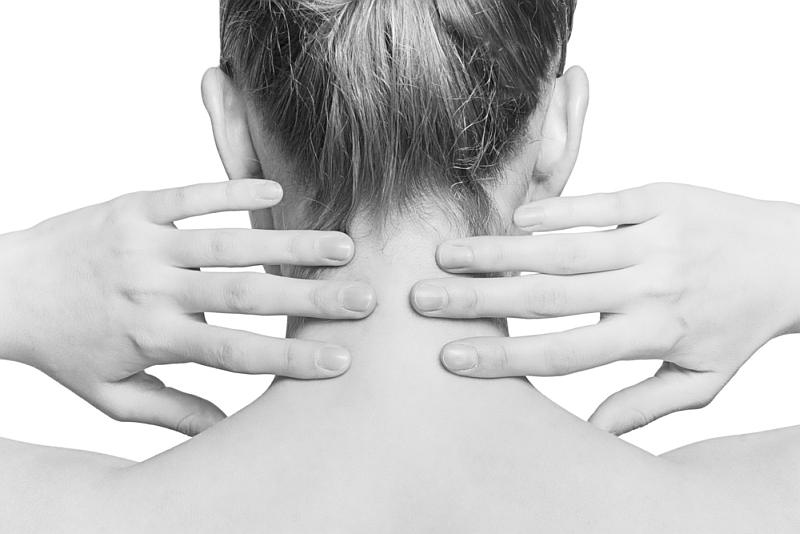
Anatomically, the neck is the region between the base of the skull and the clavicles (collar bones) that connects the head to the trunk. It contains several important blood vessels, nerves and organs. The neck also has a number of muscles and bones which help to support and protect these structures.

The bones of the neck serve to protect the spinal cord and to provide a site of attachment for the numerous neck muscles. The bones of the neck include the seven cervical vertebrae, the hyoid bone, the clavicles and the manubrium of the sternum.
Vertebrae
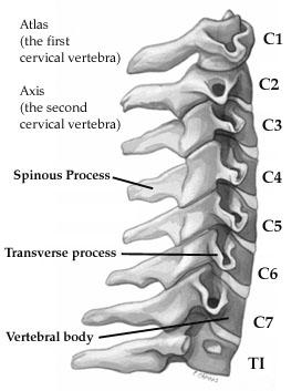

Vertebrae C1, C2 and C7 are classified as atypical vertebrae, as they have special features which are not common to the typical vertebrae (C3-C6). The different parts of the vertebrae have different functions.
The thicker, stronger, anterior bodies of the vertebrae are important for weight bearing, and thereby are important in supporting the weight of the head. The facets of the vertebrae help to join adjacent vertebrae, and the movement at these joints allows the neck to be flexible.
Typical Vertebrae – C3, C4, C5, C6
Vertebrae C3-6 all share similar features and are deemed to be typical vertebrae. The key features of these vertebrae include:
Body: The body of a vertebra is the thick, broad, anterior section. The vertebral bodies in the cervical region are rectangular. In comparison to the thoracic and lumbar vertebrae, the cervical vertebral bodies are much smaller as they do not bear as much weight.
Intervertebral discs: The intervertebral discs are located between the vertebral bodies of adjacent vertebrae and help to absorb shock. The discs in the cervical spine are thinner than in the lumbar spine; however, still thicker than the vertebral bodies they support.
Vertebral foramen: This is the hole in the vertebra between the body and the arch through which the spinal cord and meninges run. This is triangular shaped and surprisingly large in the cervical region. This is because there is a normal enlargement of the spinal cord in the cervical region (called the cervical enlargement), and is the area in which the main nerves to the arms leave the spinal cord.
Transverse processes: The transverse processes are lateral projections of the vertebrae which have facets projecting off them. There is a superior facet to articulate with the vertebra above, and an inferior facet which articulates with the vertebra below. There are a few features on the transverse process which are unique to the cervical vertebrae. Firstly, there is an oval-shaped hole called the transverse foramen, which transmits the vertebral artery and accompanying veins. The transverse processes also have two projections on them, called the anterior and posterior tubercules. These are the sites of attachment for the scalene and levator scapulae muscles.
Spinous Processes: the spinous process is a projection off the vertebra which faces posteriorly (i.e. towards the back). The spinous processes are the “bumps” which you can see often seen on the back and neck of individuals. In the cervical region, these are short (with the exception of C6 and C7) and are sometimes bifid (split into two).
Atypical Vertebrae
C1 (Atlas): C1 is a particularly unusual vertebra in that it does not have a body or a spinous process. Instead, it has anterior and posterior arches, which both have tubercules. The lateral parts of C1 are called lateral masses, which are the thickest part of the vertebrae and are involved in bearing the weight of the head. The superior surfaces of the lateral masses articulate with the occipital condyles on the base of the skull. The transverse processes of C1 arise from the lateral masses. As with the other cervical vertebrae, the transverse processes of C1 possess transverse foramina. Another feature unique to C1 is the transverse ligament, a structure which attaches to both lateral masses and holds the dens of C2 (discussed below) in place to prevent displacement of the dens into the spinal cord.
C2 (Axis): The most striking feature of C2 is the dens, a process which projects superiorly towards C1. It sits in front of the spinal cord. The dens is an important anatomical feature because it allows us to rotate our heads; it acts like a pivot. It articulates with a facet on C1 and is supported posteriorly by the transverse ligament. C2 also has prominent facets on the superior surface which articulate with C1.
C7 (Vertebra Prominens): C7 has a much longer spinous process than the typical cervical vertebrae, and often stands out as a prominent bump at the base of one’s neck when looking at someone from behind. In addition, in C7, the foramina are small or absent. When present, the C7 foramina do not transmit the vertebral arteries; instead, they only carry small accessory veins.
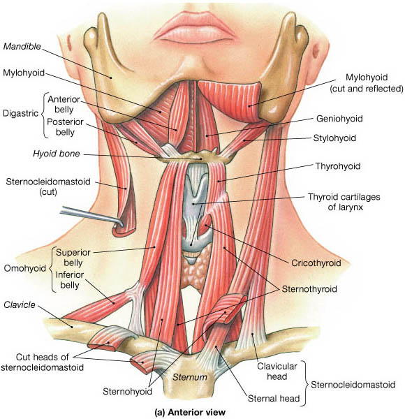
Hyoid Bone
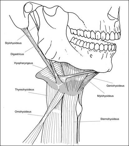

The hyoid bone is the only bone in the human body which does not directly articulate with any other bone. However; it does not merely float; there are strong muscles and ligaments which attach the hyoid to various other structures in the head and neck, including the scapulae (shoulder blades), manubrium, mandible (jaw), styloid process (part of the skull near the ear) and the thyroid cartilage of the larynx. Functionally, the hyoid is a very important bone because it helps to keep the airway open, and also serves as the attachment site for many of the neck muscles.
The hyoid is a horse-shoe shaped bone. It has a thick anterior part, which is called the body. Attaching to the sides of the hyoid body are the greater horns, which project backwards. Another smaller pair of horns, called the lesser horns, project upwards from an area close to the junction of the greater horns with the body of the hyoid bone.
Clavicle: The clavicle is colloquially referred to as the collar bone. It is a bone that is slightly curved into an S-shape and connects the upper limb to the trunk. The clavicle articulates with the manubrium at the clavicular notch. It joins to the shoulder girdle via an articulation with the acromion process of the scapula. The clavicle is important for the biomechanics of the shoulder and serves as a site of attachment for numerous muscles and blood vessels.
Manubrium of the sternum: The manubrium is the most superior part of the sternum (the breast bone). The manubrium attaches to the body of the sternum (this joint is known as the sternal angle, and is a landmark for the site of the 2nd rib), the 1st and 2nd ribs, and the two clavicles. The manubrium is also the site of attachment for some of the neck muscles. The manubrium and clavicles together form the lower border of the neck.
Summary
The bones of the head and neck play the vital role of supporting the brain, sensory organs, nerves, and blood vessels of the head and protecting these structures from mechanical damage. The 7 cervical vertebrae of the neck support the skull and organs of the head and neck. The first cervical vertebra (atlas) supports and balances the head. The second vertebra (axis) allows the head to rotate laterally to the left and the right. Spaces within the cervical vertebrae protect and conduct the spinal cord and vertebral arteries through the neck. Muscles attached tothe cervical vertebrae provide movement and posture to the head and neck.