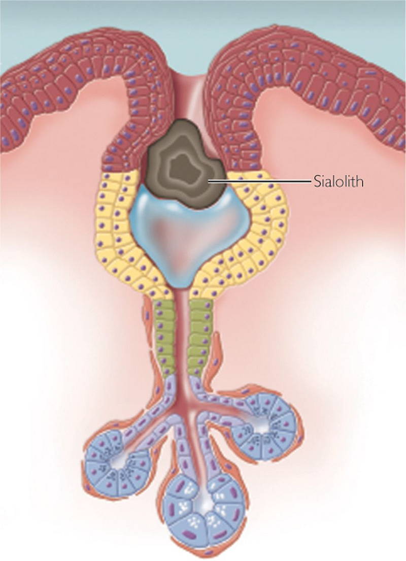
Sialolithiasis is a term used to describe stones which form in the salivary glands. The overwhelming majority of stones are found in the submandibular salivary gland, which lies near the angle of the jaw. Stones are much more likely to form in the submandibular gland because there is a greater concentration of calcium in the saliva produced here than the other glands. Its duct is also much longer than those of the other glands, which also predisposes to stone formation. They can also less commonly affect the parotid salivary gland, which lies on the face in front of the ear. It is rare for stones to affect the sublingual and other minor salivary glands.
Signs and Symptoms
Sialolithiasis is a painful condition. The pain is often made worse by eating, and the patient may notice some swelling in the gland. Some patients may also notice, or have noticed in the past, sand-like particles in their mouth. It is possible for the salivary gland to become infected, especially if the duct is blocked by the stone. In this case, there may be pus coming from the salivary gland and the mouth may also be red, tender and swollen. The patient may also feel a hard lump near their tongue if the stone is located in the end of the duct. It may also be difficult for the patient to eat if the duct is completely blocked by the stone, as saliva cannot easily enter the mouth from the affected gland.
Diagnosis
It is possible to view some salivary stones on an x-ray. This is because stones in the submandibular gland are rich in calcium phosphate. Calcium deposits are visible on an x-ray as a white mass. However, stones in the parotid glands have a different composition and are less likely to show up on x-ray. Sialography is a superior method to x-ray in detecting sialolithiasis. This is a technique which is similar to plain x-ray, however, a contrast dye is injected into the salivary gland in question which helps to better visualise the anatomy of the gland and associated structures. Ultrasounds are of little value in the diagnosis of salivary stones.
Causes
One of the main causes of salivary stones is dehydration. Dehydration reduces the water content of the saliva and causes the other components of the saliva to crystallise. A number of medications have been identified that increase the risk of developing sialolithiasis, such as certain diuretics and antidepressants.
There is also a link between Sjogren’s syndrome (an autoimmune disease) and the development of sialolithiasis. Patients with Sjogren’s syndrome have antibodies against tissue in the eyes and the salivary glands, which gradually destroy the tissue and cause symptoms of dry eyes and dry mouth. The loss of fluid content in the saliva predisposes the patient with Sjogren’s syndrome to suffer with sialolithiasis. Interestingly, gout may also cause sialolithiaisis in rare instances. However, in patients with gout-induced sialolithiasis, the crystals are made up of uric acid; the substance forming the crystals which affect the joints of the sufferer. People who suffer from chronic inflammation of the salivary glands are also more likely to develop salivary stones.
Complications
Untreated sialolithiasis can cause recurrent, painful infections, which can lead to scarring and abscess formation in the salivary gland. There are also risks involved with the treatment options for sialolithiasis. Surgical approaches to the salivary glands may result in damage to nearby nerves and blood vessels. The parotid gland is particularly at risk because the facial nerve passes through it. Intra-oral extraction techniques may also result in scarring to the duct and affect the drainage of saliva into the mouth. There is also a risk of infection following removal of the stones, due to the high volumes of bacteria in the mouth and surrounds.
Treatment
There are a number of treatment options available for sialolithiasis, largely dependent on the size and location of the stone. If the stones are small and near the end of the duct of the salivary gland, it may be possible to extract the stone via the mouth under local anaesthesia. Some larger stones may also be able to be extracted by placing a cannula in the duct and making a small cut in the duct, which allows for removal of the stone. In cases of very large, complicated stones, the entire salivary gland may be removed. Recent advances have seen the development of endoscopic techniques which can help to remove salivary stones in a much less invasive manner than other surgical methods.
If you have questions about sialolithiasis and salivary gland problems contact your local doctor, who will arrange for you to see an Ear Nose Throat Specialist. We‘ll provide you with a straightforward, efficient and very effective treatment plan targeted to your condition.


